MENU

DriveAFM-FluidFM-brand-angle

FluidFM®: Fluidic force microscopy

The established microfluidic tool for nanomanipulation and single-cell biology

FluidFM literally brings microfluidics to the tip of an AFM cantilever. This way, it combines the microfluidics with the force sensitivity and positional accuracy of an AFM allowing a range of exciting applications in single-cell biology and nanoscience.

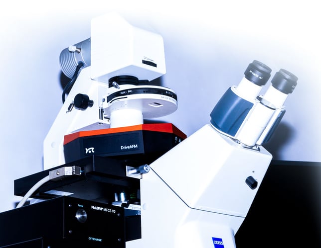
Nanosurf has a long experience providing AFM systems with FluidFM having launched the FlexAFM with FluidFM in 2011 together with Cytosurge AG. This integration of FluidFM on Nanosurf platforms has grown in time and now the FluidFM technology available for DriveAFM, FlexAFM, and CoreAFM platforms.

How does FluidFM work?

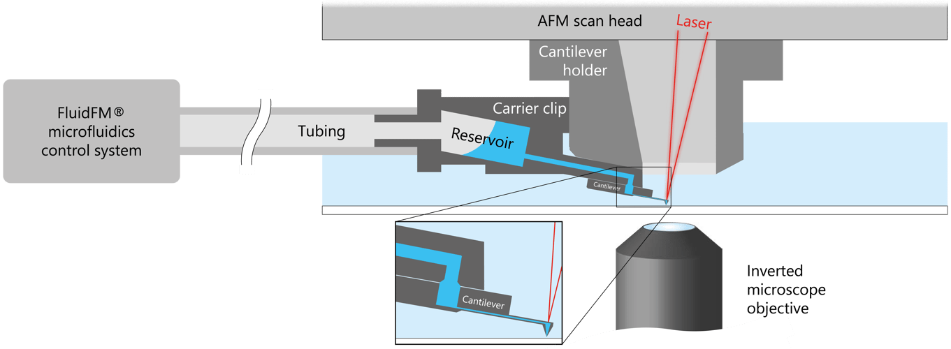
At the heart of the technology lies a hollow cantilever with an aperture at the tip, through which fluids are delivered or withdrawn. Liquid is filled into a small reservoir attached to the cantilever. Air pressure is applied to the other side of the reservoir through tubing, propelling the liquid through the channel in the cantilever and out of the aperture at the tip. By controlling the pressure and the flow rate of the liquid, FluidFM can be used to perform a wide range of tasks, including printing, dispensing, and probing.

The AFM component of FluidFM allows for precise positioning and force measurements at the nanoscale level. The microfluidic component allows for precise control of the amount and location of the liquid being delivered or withdrawn.

FluidFM cytoclip schematic

The combination of these capabilities allow for the manipulation of individual cells, bacteria, or other biological structures, as well as to deposit and remove materials with high accuracy, making it a powerful tool for a variety of applications in life sciences, materials science, and nanotechnology.

FluidFM application areas

With FluidFM you can perform a wide range of experiments

FluidFM-icon-single-cell-adhesion

Single cell adhesion

FluidFM-icon-colloidal-probe

Colloidal spectroscopy

FluidFM-icon-single-cell-injection

Single cell injection

FluidFM-icon-spotting

Spotting

FluidFM-icon-bacterial-adhesion

Single bacterium adhesion

FluidFM-icon-single-cell-isolation

Single cell isolation

FluidFM-icon-single-cell-extraction

Single cell extraction

FluidFM-icon-nanolithography

Nanolithography

FluidFM-icon-picobalance

PicoBalance

FluidFM-icon-SICM

SICM

Different applications require different FluidFM probes

  • FluidFM micropipettes: tipless cantilevers with opening at the cantilever end
  • FluidFM nanopipettes: cantilevers with opening at the tip apex
  • FluidFM nanosyringes: cantilevers with opening at the side of the tip, maintaining a sharp apex to penetrate biological membranes for injection and extraction

Highly accurate pressure, force, and position control with optical sample access

  • From add-on to fully integrated system in the DriveAFM Studio software
  • Fast and accurate FluidFM microfluidics control system
  • Integrates with optical microscopy techniques, e.g. fluorescence
  • Highest force resolution and up to 20 µm z-range internally
ir_attachment_1394_jpeg_rgb
fpm-sorted-cells

Optical cell selection by fluorescence properties (left image). The cantilever was gently placed on the cell under force control and trypsin administration was monitored from a co-deposited fluorescence dye (blue). After release, the cell was picked up with the same cantilever and isolated from the rest by placing it in a separate well, sorted by fluorescence (right image; adapted from Lab Chip (2014) 14, 402‑414 with permission from The Royal Society of Chemistry). Data courtesy: O. Guilaume-Gentil, ETH Zürich, Switzerland.

A tool for research pioneers

  • FluidFM enables original research at the frontiers of science from bacterial adhesion to cell-cell interaction and from spotting to injection and extraction
  • New: FluidFM can be combined with PicoBalance to measure the mass of cells and microparticles
FluidFM-mass-measurements-picobalance

Left: Optical microscope image of FluidFM micropipettes with differently sized colloidal particles attached.
Right: PicoBalance mass measurement results for 7 different beads.

An integrated solution with optical, force, and fluidic control

FluidFM probes

All FluidFM probes come in sterile blister packs and pre-mounted on plastic carrier clips. Each blister pack has a QR code that can be read with the QR code reader supplied with the FluidFM system.

Cantilevers are easily mountable - even when wearing gloves.


FluidFM_cantilever_holder_1000x600

Basic integration with FlexAFM and CoreAFM

FluidFM can be operated with basic integration for FlexAFM and CoreAFM, providing microfluidics control during the contact phase in spectroscopy for adhesion, spotting, injection or extraction experiments.


FluidFM_Flex_spotting_1000x600

Perfect integration with DriveAFM and Nanosurf Studio

In the Studio software platform, FluidFM is seamlessly integrated. Besides access to the pressure controller during spectroscopy, Viewport allows targeting of cells or areas of interest directly in live feed of the optical camera. Furthermore DriveAFM uniquely provides the PicoBalance functionality: measure the mass of microparticles or single cells.

FluidFM_studio_viewport_1000x600
Zambelli

Prof. Tomaso Zambelli, Laboratory of Biosensors and Bioelectronics, ETH Zurich

FluidFM at ETH Zurich

At the Laboratory of Biosensors and Bioelectronics (ETH Zurich) we have been using the FlexAFM system with FluidFM add-on since the very early days of FluidFM. Together with Cytosurge, SmartTip and Nanosurf we developed this technology establishing several applications from cell adhesion, colloidal spectroscopy, force-controlled SICM and patch clamp, to local chemical cell stimulation. On the other hand, together with the Institute of Microbiology (ETH Zurich, Prof. Vorholt) we pioneered single-cell protocols like injection, extraction and isolation. Our results are well-received by the scientific community because of their originality stimulating ideas for experiments never attempted before. In this groundbreaking phase the Nanosurf team constantly supported us with interest, competence, and flexibility relying on their long-dated AFM experience.


FluidFM in action

Single cell injection of DAPI into the cytoplasm between two living Fibroblast cells. Performed with FluidFM integration on DriveAFM.

Example of single cell force spectroscopy: in this video a single cell is detached from fully adherent and confluent culture. The measured forces both depend on the substrate as well as the bonds to the neighboring cells. Courtesy of A. Sancho and J. Groll, Functional Materials for Medicine and Dentistry, University Hospital of Würzburg.

Working principle of single cell injection explained: Single cell nano-injection enables targeted introduction of foreign materials into a specific cell. Stimulating selected cells within a colony with your desired drugs or gene vectors provides new insights for life science, biology and medicine.

Example of nanolithography by FluidFM spotting. Droplets of paraffin oil are deposited on a glass substrate with a FluidFM nanopipette. Distance between spots down to approx. 5 µm in this spiral.

FluidFM is available on Nanosurf research AFM systems

FluidFM is available on Nanosurf's  DriveAFM, FlexAFM and CoreAFM systems. Contact us to identify the right setup for your research requirements.

DriveAFM-FluidFM_800px

DriveAFM

The high-end solution for FluidFM

FluidFM-FlexAFM_800px

FlexAFM

The most published FluidFM solution

CoreAFM-FluidFM

CoreAFM

The most affordable FluidFM solution

Download the FluidFM brochure

FluidFM-brochure_thumbnail

Publications


Nature 2022/08/17

Live-seq enables temporal transcriptomic recording of single cells

Wanze Chen, Orane Guillaume-Gentil, Pernille Yde Rainer, Christoph G. Gäbelein, Wouter Saelens, Vincent Gardeux, Amanda Klaeger, Riccardo Dainese, Magda Zachara, Tomaso Zambelli, Julia A. Vorholt & Bart Deplancke

Single-cell transcriptomics (scRNA-seq) has greatly advanced our ability to characterize cellular heterogeneity. However, scRNA-seq requires lysing cells, which impedes further molecular or functional analyses on the same cells. Here, we established Live-seq, a single-cell transcriptome profiling approach that preserves cell viability during RNA extraction using fluidic force microscopy, thus allowing to couple a cell’s ground-state transcriptome to its downstream molecular or phenotypic behaviour. To benchmark Live-seq, we used cell growth, functional responses and whole-cell transcriptome read-outs to demonstrate that Live-seq can accurately stratify diverse cell types and states without inducing major cellular perturbations. As a proof of concept, we show that Live-seq can be used to directly map a cell’s trajectory by sequentially profiling the transcriptomes of individual macrophages before and after lipopolysaccharide (LPS) stimulation, and of adipose stromal cells pre- and post-differentiation. In addition, we demonstrate that Live-seq can function as a transcriptomic recorder by preregistering the transcriptomes of individual macrophages that were subsequently monitored by time-lapse imaging after LPS exposure. This enabled the unsupervised, genome-wide ranking of genes on the basis of their ability to affect macrophage LPS response heterogeneity, revealing basal Nfkbia expression level and cell cycle state as important phenotypic determinants, which we experimentally validated. Thus, Live-seq can address a broad range of biological questions by transforming scRNA-seq from an end-point to a temporal analysis approach.


Bioengineering 20229(10), 567

Adhesion Forces of Oral Bacteria to Titanium and the Correlation with Biophysical Cellular Characteristics

Katharina Doll-Nikutta, Andreas Winkel, Ines Yang, Anna Josefine Grote, Nils Meier, Mosaieb Habib, Henning Menzel, Peter Behrens and Meike Stiesch

Bacterial adhesion to dental implants is the onset for the development of pathological biofilms. Reliable characterization of this initial process is the basis towards the development of anti-biofilm strategies. In the present study, single-cell force spectroscopy (SCFS), by means of an atomic force microscope connected to a microfluidic pressure control system (FluidFM), was used to comparably measure adhesion forces of different oral bacteria within a similar experimental setup to the common implant material titanium. The bacteria selected belong to different ecological niches in oral biofilms: the commensal pioneers Streptococcus oralis and Actinomyces naeslundii; secondary colonizer Veillonella dispar; and the late colonizing pathogens Porphyromonas gingivalis as well as fimbriated and non-fimbriated Aggregatibacter actinomycetemcomitans. The results showed highest values for early colonizing pioneer species, strengthening the link between adhesion forces and bacteria’s role in oral biofilm development. Additionally, the correlation between biophysical cellular characteristics and SCFS results across species was analyzed. Here, distinct correlations between electrostatically driven maximum adhesion force, bacterial surface elasticity and surface charge as well as single-molecule attachment points, stretching capability and metabolic activity, could be identified. Therefore, this study provides a step towards the detailed understanding of oral bacteria initial adhesion and could support the development of infection-resistant implant materials in future.


Cell 166 (2016) doi: 10.1016/j.cell.2016.06.025

Tunable Single-Cell Extraction for Molecular Analyses

O Guillaume-Gentil, RV Grindberg, R Kooger, L Dorwling-Carter, V Martinez, D Ossola, M Pilhofer, T Zambelli, and JA Vorholt

Researchers from ETH Zurich have published extraction of sub-picoliter samples of nucleoplasm and cytoplasm from live cells in the renowned journal Cell. Measurements were performed with the Nanosurf Flex-FPM system that allowed single-cell extraction without killing the cells. The paper describes the method of single-cell extraction as well as the molecular analysis of the extracted samples by TEM, protein assays and PCR. The results illustrate a new FluidFM® application to study cell processes at the single-cell level thus enabling studying the heterogeneity of cells.


ChemPhysChem (2017) doi: 10.1002/cphc.201700780

Local Chemical Stimulation of Neurons with the Fluidic Force Microscope (FluidFM)

Mathias J. Aebersold, Harald Dermutz, László Demkó, José F. Saenz Cogollo, Shiang-Chi Lin, Conrad Burchert, Moritz Schneider, Doris Ling, Csaba Forró, Hana Han, Tomaso Zambelli and János Vörös

This new stimulation approach, which combines FluidFM for gentle and precise positioning with a microelectrode array read-out, makes it possible to modulate the activity of individual neurons chemically and simultaneously record their induced activity across the entire neuronal network. The presented platform not only offers a more physiological alternative compared with electrical stimulation, but also provides the possibility to study the effects of the local application of neuromodulators and other drugs.


Microscopy & Analysis, June 2018

FluidFM: Precise fluidic positioning and delivery platform with applications in cell biology and soft matter

Patrick Frederix, Paul Werten, Dalia Yablon

The platform of fluid force microscopy (FluidFM) offers researchers unique capabilities in precise and well-controlled fluid delivery in small quantities down to femtoliters to the surface. It is based on a microfluidic controller coupled with an innovative AFM probe where a microchannel is hollowed out so that the probe essentially functions as a nanopipette. Applications of FluidFM are described including enhanced manipulation of cells, improved force measurements on biological materials including bacteria, and fluidic writing on hydrogels.


J. Phys. Chem. C 120 (2016) 18015–18027

Colloidal Properties of Recombinant Spider Silk Protein Particles

N Helfricht, E Doblhofer, JFL Duval, T Scheibel, and G Papastavrou

Researchers from the group of Prof. Georg Papastavrou at the University of Bayreuth used the Nanosurf Flex-FPM system to perform direct force measurements in the sphere/sphere geometry. Colloidal particles have been prepared from polyanionic and polycationic recombinant spider silk proteins. The amino acid sequences of these spider silk proteins were identical except for 16 residues bearing either cationic or anionic groups, leading to opposite surface charges. Electrokinetic measurements and modelling predicted a soft and porous structure of these protein particles. The presence of a fuzzy, ion-permeable interface has been confirmed by direct force measurements with colloidal particles aspirated reversibly to a FluidFM® probe.