The extraordinarily successful launch of the DriveAFM has been the crowning achievement for the hard work of Nanosurf's development team over the past years. We are pleased to see the overwhelming market response to its performance and versatility. This positive feedback gives us unfettered motivation to keep enhancing the abilities of the instrument. We are looking along an exciting R&D pipeline, bursting with new ideas and features, and are currently focussing on further improving the ease of use and speed of the DriveAFM.
As we prioritized the functionality for life science initially, the past months have been focused on making a spot landing in the field of materials research: the latest addition to our nano-electrical characterization toolbox is a new scanning microwave microscopy solution. It has been developed together with the Swiss Federal Institute of Metrology (METAS) and is compatible with all of our research grade instruments (CoreAFM, FlexAFM, and DriveAFM).
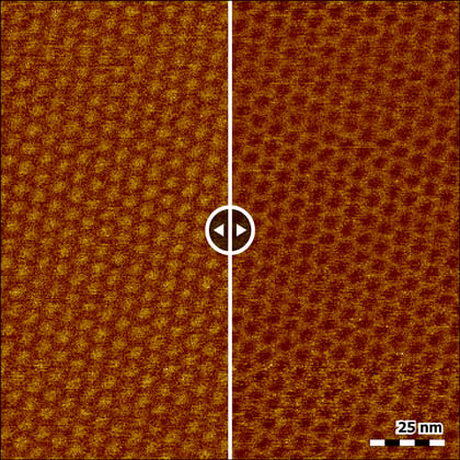
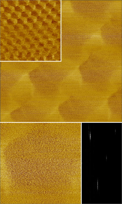
To top this off, our application team made some breakthrough findings for graphene in collaboration with scientists in the field, as well as showing clear visualizations of moiré superlattices, measured by PFM on a FlexAFM (in a glovebox) .
Stay tuned for more news on the latest developments.
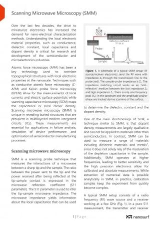
SMM is a scanning probe technique that measures the interactions of a microwave between a sharp tip and the sample. The ratio between the power sent to the tip and the power received after being reflected at the tip-sample contact can be used to determine the dielectric constant and the dopant density. SMM is currently available on FlexAFM and CoreAFM, and will soon be available on DriveAFM as well.
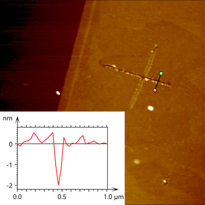
During an inspiring visit to the Kim group at Harvard University, Dr. Ed Nelson tested graphene cutting with the FlexAFM. It turned out to be astonishingly easy, and he saved some nice images of a cut cross.
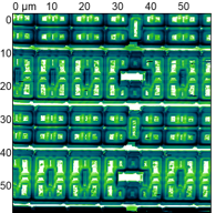
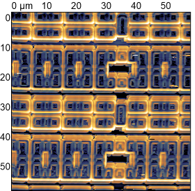
Dr. Patrick Frederix measured moiré patterns with a CoreAFM, a FlexAFM and a DriveAFM on samples received from Nanoelectronics group TIFR, India. “These findings prove the power of Nanosurf instruments as a tool for graphene research” says Dr. Frederix.
As already shown in the literature, the moiré superlattice measured by PFM also causes a shift in contact resonance, which can explain the phase and amplitude contrast. This would imply that the moiré pattern should also be visible when measured by other means of excitation on the resonance peak, like force modulation. This was successfully verified on DriveAFM, using photothermal excitation to oscillate the cantilever at the contact resonance.
"The icing on the cake was the visualization of both the Moiré superlattice and atomic lattice of graphene in a single image" was Dr. Frederix' satisfied reaction after assessing the data.
Has this piqued your interest?
Keep both eyes open for a summary of applications of AFM as a tool for graphene research in our new application note, which we will be publishing later this week.
Recent application notes
Scanning Microwave Microscopy (SMM)
SMM is a scanning probe technique that measures the interactions of a microwave between a sharp tip and the sample. This technique can yield information about the local capacitance from a range of materials including metals, semiconductors and dielectrics.
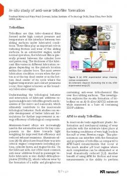
Tribofilms are thin tribochemical films formed under high contact pressure and temperature at the interface between two sliding surfaces under lubricated conditions.
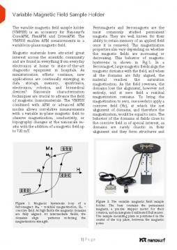
The variable magnetic field sample holder (VMFSH) is an accessory for Nanosurf’s CoreAFM, FlexAFM and DriveAFM. The VMFSH enables AFM measurements in a variable in-plane magnetic field of up to 720 mT.
Nanoscale Mapping using VertiSense™ Scanning Thermal Microscopy
Ami Chand, founder of Appnano, will present details of a new thermal Probe that provides sensitive and direct local thermometry data. The webinar will cover details of the SThM module, the structural details of this unique SThM probe, and review various applications.
In case you missed any of our previous webinars, you can view a recording on our video page.
Recent installations
FlexAFM installed at Roskilde University
A FlexAFM with DIMO option was installed at the Department of Science and Environment at Roskilde University in Denmark. The Digital Inverted Microscope Option (DIMO) allows the system to be used easily for analysis of both materials and life science samples. The FlexAFM will be used by a number of research groups to study nano-scale self-organized structures that are central building blocks in both nanotechnological devices and in many biological systems and materials.
Alphacen installed at Newport MKS
We recently installed a customized large stage Alphacen AFM at Newport MKS. This AFM complements Newport’s metrology lab and enables sub-Angstrom roughness measurements on samples that come in a variety of shapes and sizes. We look forward to working with Newport to further develop unique tools and provide them with support they need to achieve their goals.
FlexAFM installed at NIT, Surathkal, India
A FlexAFM was installed at the Central Research Facility, National Institute of Technology, Surathkal India. The AFM will be housed in a central facility and will be used to study various materials including biomaterials and semiconductors. The FlexAFM will also be made available to other nearby research institutes as a service. Dr. Sathyanarayana’s group from the Dept. of Physics will be the primary users of the AFM.
Evaluation of Streptococcus mutans Adhesion to Stainless Steel Surfaces Modified Using Different Topographies Following a Biomimetic Approach. https://doi.org/10.3390/coatings11070829